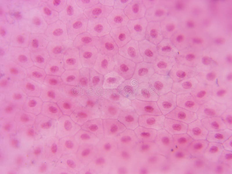
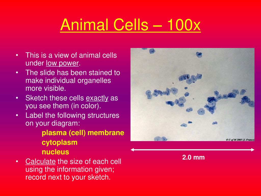
animal cell under microscope 100x
Scanning probe microscopy SPM is a branch of microscopy that forms images of surfaces using a physical probe that scans the specimen. They play a major role in ensuring clear sharp images are produced with a high magnification of 400X and above.
Typical Animal Cell Center 400x Stock Image Image Of Visible Compound 152965979
Magnification is the ratio of enlargement or reduction of an image.

. For experiments U251-RedFLuc cells were trypsinized as described above. SPM was founded in 1981 with the invention of the scanning tunneling microscope an instrument for imaging surfaces at the atomic levelThe first successful scanning tunneling microscope experiment was done by Gerd Binnig and. Must contain at least 4 different symbols.
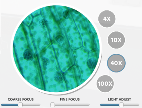
The wildly grown leaves of D. Condensers are lenses that are used to collect and focus light from the illuminator into the specimen. To calculate the total magnification you need to take the power of the objective lens 10X 40X 100X and multiply it by the power of the eyepiece 10X.
ELx405 Select Deep Well Microplate Washer is a robot compatible full plate washer incorporating BioTeks Dual-Action manifold with independent filling and evacuation control for precise overfill washing and overflow protection. Magnification is usually expressed as multiples of a number for example X10 X40 X100 etc. Iodine crystal violet and methylene blue are examples of simple stains.
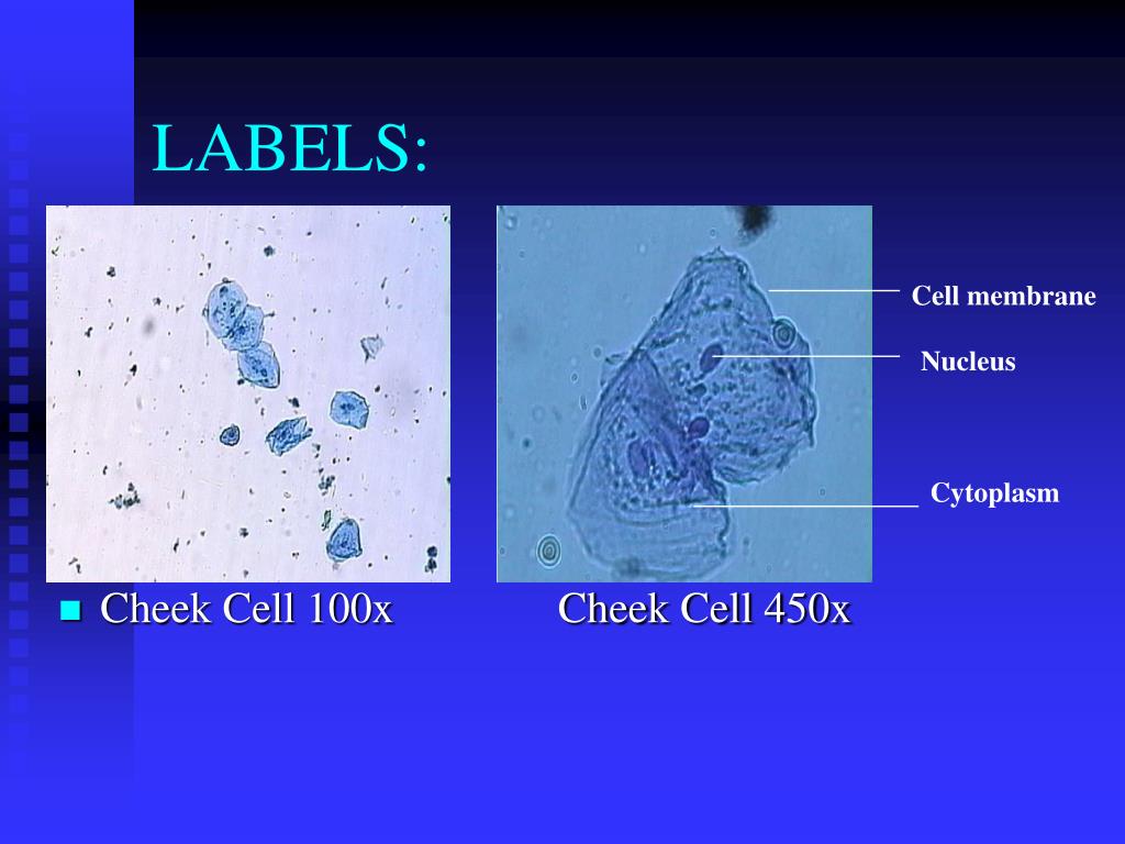
In addition the electron microscope is required to resolve the structure of mitochondria bacteria viruses and large protein complexes. ASCII characters only characters found on a standard US keyboard. Under the microscope animal cells appear different based on the type of the cell.
This is usually smaller than a lake and may either be man-made or natural. The compound microscope typically has three or four magnifications - 40x 100x 400x and sometimes 1000x. At 100x magnification you will be able to see 2mm.
Transcription factors are known to cooperate with each other in their interaction with cognate DNA binding motifs. Linearis were collected around the area of Serdang Selangor Malaysia between February and March 2012. 驚きの吸収力とずっとふっくらなタオルThe Last Towelラストタオル最大の特徴は 話題の水に溶ける魔法の糸スーパーゼロ触れただけで水を吸い取る圧倒的吸水力を実感ください.
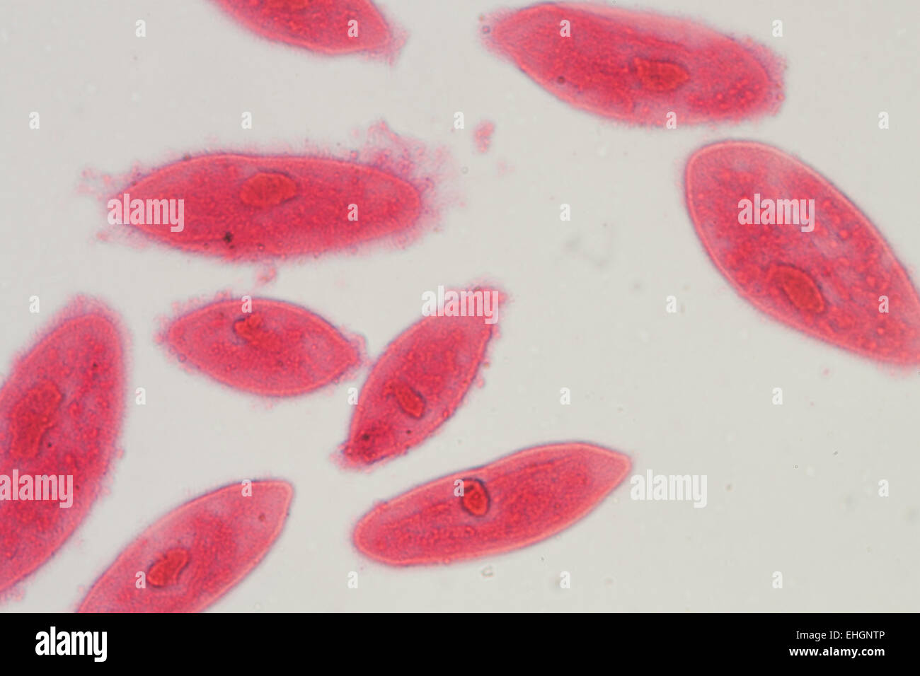
Z-stack of embryo tail was performed through approximately 40μm of tissue obtaining a z-slice every 3μm. Pond Water Under the Microscope. Under a higher magnification of 100X nuclei of the cells appear.
At 100x magnification you will be able to see 2mm. Report a superfamily of 30 factors that recognizes overlapping GC-rich sequences in mammalian genomes and that. To see the cell organelles you will need to get a higher magnification usually with a 40x-100x objective lens.
Successful transduction was confirmed with in vitro bioluminescence imaging as described below. The images of Paulownia wood hair and frogs blood were captured with a high power compound microscope using a Nikon camera adapter. Proliferation and appearance of cell cultures were assessed with a Telaval 31 light microscope 100x magnification.
Plant Material and Preparation of Methanol Extract D. Or total mTECs by crossing mice expressing Cre recombinase under the Foxj1 Ckm or Foxn1 promoters with Rosa26-LSL-eYFP reporter mice hereafter referred to as. Advanced Fully Multi-Coated lenses and powerful BAK4 Roof Prism provide sharpness and exceptionally accurate colors throughout the whole light spectrum.
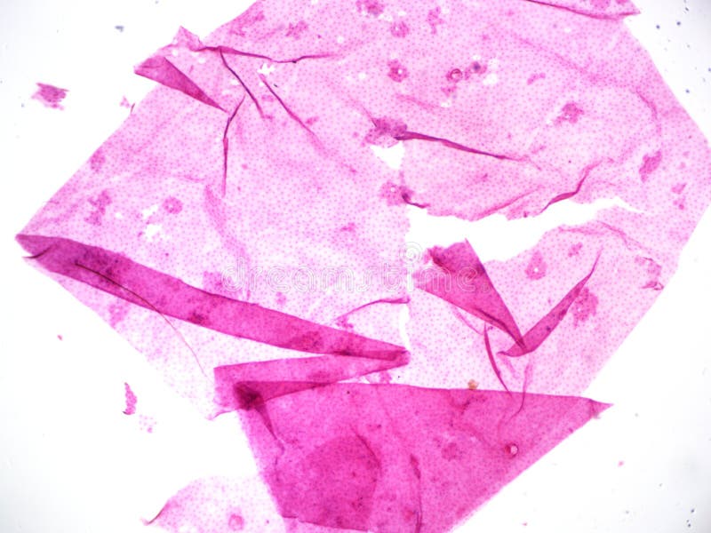
Animal cell under the microscope. Microscope cell staining is a technique used to improve the visibility of cells and cell parts under a microscope. Video was acquired on a Nikon A1 confocal microscope using a 60x objective.
Simple epithelium is composed of a single layer of cells while stratified epithelium contains several layers. They are found under the stage next to the diaphragm of the microscope. The cell remains in prometaphase for the duration of the two-hour acquisition.
This slide showing a cross section of the mammalian trachea wind pipe contains. High refractive index lens immersion. Resolution is the ability of the lens to show the fine details of.
Explore topics on usage care terminology and then interact with a fully functional virtual microscope. This work is licensed under a Creative Commons Attribution-NonCommercial-NoDerivs 25 LicenseCreative Commons Attribution-NonCommercial-NoDerivs 25 License. NEB is inferred by the irregular appearance of the nucleus.
While some can be seen with the naked eye others are too small and will require the use of a microscope to be able to properly observe. At 400x magnification you will be able to see 045mm or 450 microns. Pond water contains a variety of plant and animal life.
Lesson Description BioNetworks Virtual Microscope is the first fully interactive 3D scope - its a great practice tool to prepare you for working in a science lab. Available low-flow rates and angled dispensing make the ELx405 particularly useful in cell-based assays. Pond water refers to a standing body of water.
When you are ready challenge your knowledge in the testing section to see what you have learned. A nucleus or a cell wall can be seen more clearly by using different stains. At 40x magnification you will be able to see 5mm.
Clear High Definition Magnification up to 100x Experience crystal clear vision with Comfortable eyecups long eye relief for a perfect fit with or without glasses. A typical animal cell is 1020 μm in diameter which is about one-fifth the size of the smallest particle visible to the naked eye. The leaves have been previously identified and a voucher specimen SK 198711 has been deposited at the Herbarium of the Institute of Bioscience.
5501470 Zeiss White Plains NY USA. Make a wet or dry mount with a coverslip. Medullary thymic epithelial cells repurpose the lineage-defining transcription factors of diverse extra-thymic cell types to create cellular mimics of the peripheral self within.
Epithelial tissues are classified according to the number of cell layers that make up the tissue and the shape of the cells. 6 to 30 characters long. This method requires a very high numerical aperture objective at least 14 but preferably 145 to 16 and partial illumination of the microscope field from one side by a small sport or more uniform illumination by a thin annulus.
At 1000x magnification you will be able to see 0180mm or 180 microns. Nikon offers 60x and 100x TIRF objectives with numerical aperture 149. Differentiate between a condenser and an Abbe condenser.
You can see a variety of cells pretty well with the light microscope.
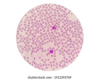
Lemur Blood Smear Under 100x Light Stock Photo 1912293769 Shutterstock
![]()
Typical Plant Cell 100x Magnification Stock Image Image Of Cells Magnification 152965909
Solved Bio 101 Lab 03 Microscopy And Cells Notification Objectives Course Hero
Animal Cell Microscope Hi Res Stock Photography And Images Alamy
Typical Animal Cell Center 40x Stock Image Image Of Typical Science 152965947

Blood Smear Show Platelet Increaseplatelet More Than 25 Cells Per 100x Microscope Stock Photo Download Image Now Istock
180 100x Magnification Stock Photos Pictures Royalty Free Images Istock
Virtual Microscope Animal And Plant Cell Tutorial Ppt Download
Virtual Microscope
Ppt Post Lab Plant Animal Cells Or Powerpoint Presentation Free Download Id 5665034

Microscopic View Of Algal Cells Cultured With And Without Co 2 Under Download Scientific Diagram
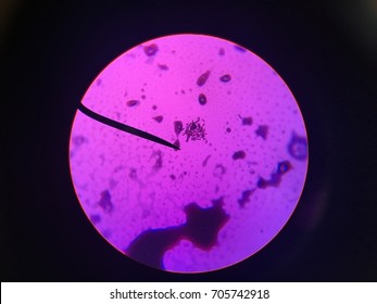
Coliforms Under Light Microscope 100x Stock Photo 705742918 Shutterstock
Solved Bio 101 Lab 03 Microscopy And Cells Notification Objectives Course Hero

509 Animal Cell Structure Photos And Premium High Res Pictures Getty Images
![]()
Typical Plant Cell 100x Magnification Stock Image Image Of School College 152965951

180 100x Magnification Stock Photos Pictures Royalty Free Images Istock
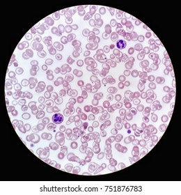
Human Blood Smear Under 100x Light Stock Photo 751876783 Shutterstock
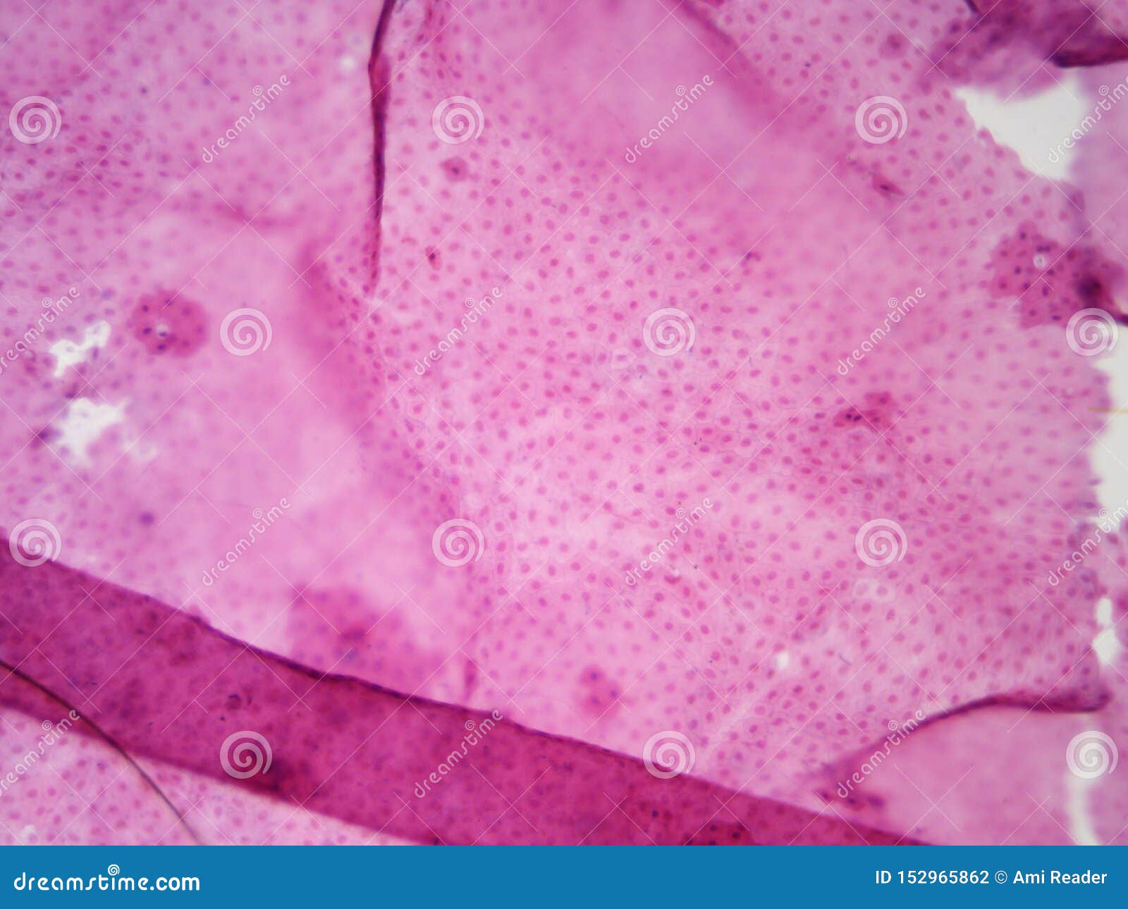
Typical Animal Cell Center 100x Stock Photo Image Of 100x School 152965862
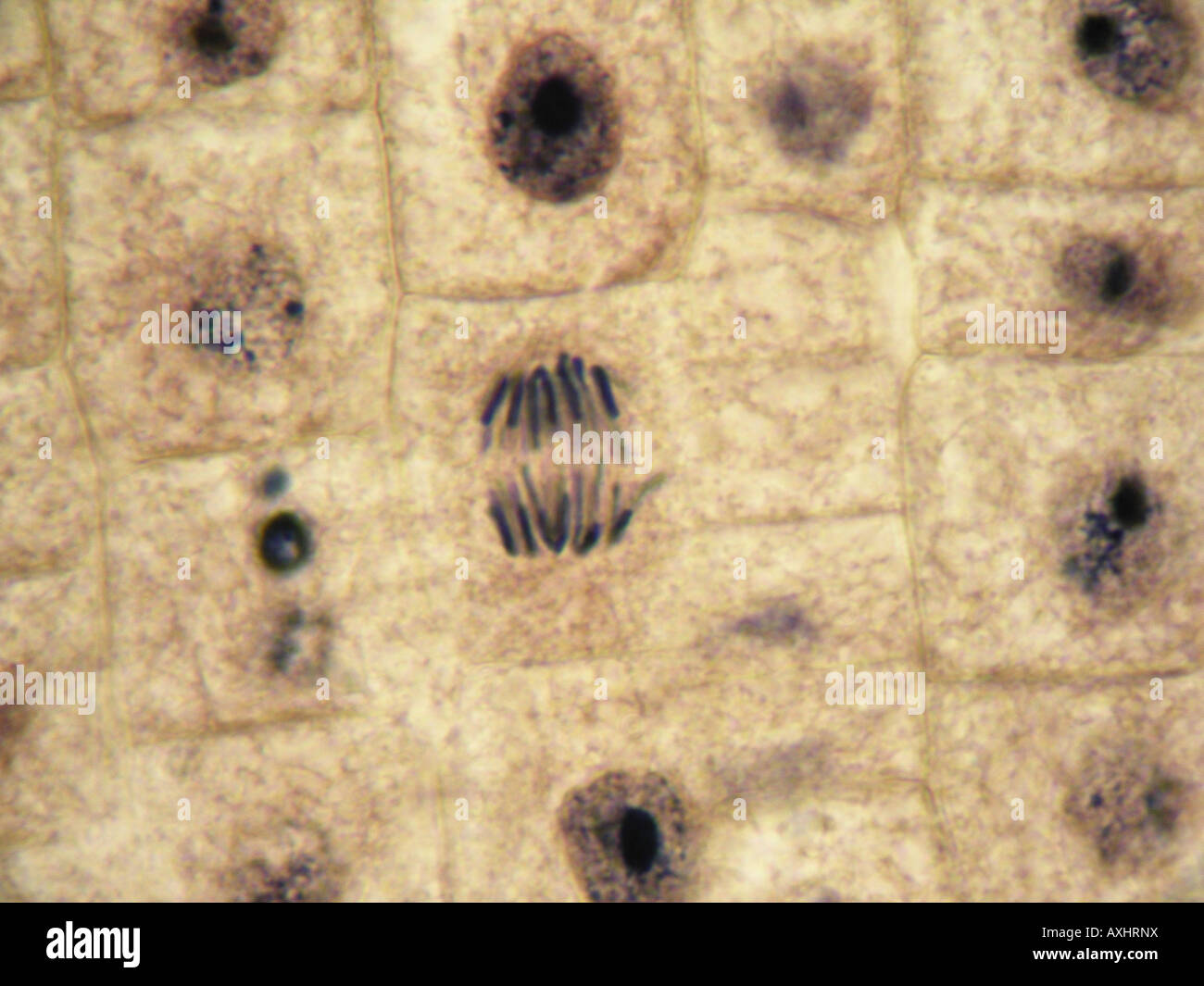
Mitosis Anaphase In Onion Tissue At 1000x Under Optical Microscope Inmersion Oil Objetive 100x Stock Photo Alamy